A | B | C | D | E | F | G | H | CH | I | J | K | L | M | N | O | P | Q | R | S | T | U | V | W | X | Y | Z | 0 | 1 | 2 | 3 | 4 | 5 | 6 | 7 | 8 | 9
| Bryozoa Temporal range: Contested Cambrian records (Pywackia, Protomelission[3])
| |
|---|---|

| |
| "Bryozoa", from Ernst Haeckel's Kunstformen der Natur, 1904 | |
| Scientific classification | |
| Domain: | Eukaryota |
| Kingdom: | Animalia |
| Clade: | Lophophorata |
| Phylum: | Bryozoa Ehrenberg, 1831[4] |
| Classes | |
| Synonyms[5] | |
|
Ectoprocta (Nitsche, 1869) (formerly subphylum of Bryozoa) | |
Bryozoa (also known as the Polyzoa, Ectoprocta or commonly as moss animals)[6] are a phylum of simple, aquatic invertebrate animals, nearly all living in sedentary colonies. Typically about 0.5 millimetres (1⁄64 in) long, they have a special feeding structure called a lophophore, a "crown" of tentacles used for filter feeding. Most marine bryozoans live in tropical waters, but a few are found in oceanic trenches and polar waters. The bryozoans are classified as the marine bryozoans (Stenolaemata), freshwater bryozoans (Phylactolaemata), and mostly-marine bryozoans (Gymnolaemata), a few members of which prefer brackish water. 5,869 living species are known.[7] Originally all of the crown group Bryozoa were colonial, but as an adaptation to a mesopsammal (interstitial spaces in marine sand) life or to deep‐sea habitats, secondarily solitary forms have since evolved. Solitary species has been described in four genera; Aethozooides, Aethozoon, Franzenella and Monobryozoon). The latter having a statocyst‐like organ with a supposed excretory function.[8][9]
The terms Polyzoa and Bryozoa were introduced in 1830 and 1831, respectively.[10][11] Soon after it was named, another group of animals was discovered whose filtering mechanism looked similar, so it was included in Bryozoa until 1869, when the two groups were noted to be very different internally. The new group was given the name "Entoprocta", while the original Bryozoa were called "Ectoprocta". Disagreements about terminology persisted well into the 20th century, but "Bryozoa" is now the generally accepted term.[12][13]
Colonies take a variety of forms, including fans, bushes and sheets. Single animals, called zooids, live throughout the colony and are not fully independent. These individuals can have unique and diverse functions. All colonies have "autozooids", which are responsible for feeding, excretion, and supplying nutrients to the colony through diverse channels. Some classes have specialist zooids like hatcheries for fertilized eggs, colonial defence structures, and root-like attachment structures. Cheilostomata is the most diverse order of bryozoan, possibly because its members have the widest range of specialist zooids. They have mineralized exoskeletons and form single-layered sheets which encrust over surfaces, and some colonies can creep very slowly by using spiny defensive zooids as legs.
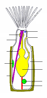
Each zooid consists of a "cystid", which provides the body wall and produces the exoskeleton, and a "polypide", which holds the organs. Zooids have no special excretory organs, and autozooids' polypides are scrapped when they become overloaded with waste products; usually the body wall then grows a replacement polypide. Their gut is U-shaped, with the mouth inside the crown of tentacles and the anus outside it. Zooids of all the freshwater species are simultaneous hermaphrodites. Although those of many marine species function first as males and then as females, their colonies always contain a combination of zooids that are in their male and female stages. All species emit sperm into the water. Some also release ova into the water, while others capture sperm via their tentacles to fertilize their ova internally. In some species the larvae have large yolks, go to feed, and quickly settle on a surface. Others produce larvae that have little yolk but swim and feed for a few days before settling. After settling, all larvae undergo a radical metamorphosis that destroys and rebuilds almost all the internal tissues. Freshwater species also produce statoblasts that lie dormant until conditions are favorable, which enables a colony's lineage to survive even if severe conditions kill the mother colony.
Predators of marine bryozoans include sea slugs (nudibranchs), fish, sea urchins, pycnogonids, crustaceans, mites and starfish. Freshwater bryozoans are preyed on by snails, insects, and fish. In Thailand, many populations of one freshwater species have been wiped out by an introduced species of snail.[14] A fast-growing invasive bryozoan[which?] off the northeast and northwest coasts of the US has reduced kelp forests so much that it has affected local fish and invertebrate populations[citation needed]. Bryozoans have spread diseases to fish farms and fishermen. Chemicals extracted from a marine bryozoan species have been investigated for treatment of cancer and Alzheimer's disease, but analyses have not been encouraging.[15]
Mineralized skeletons of bryozoans first appear in rocks from the Early Ordovician period,[1] making it the last major phylum to appear in the fossil record. This has led researchers to suspect that bryozoans arose earlier but were initially unmineralized, and may have differed significantly from fossilized and modern forms. In 2021, some research suggested Protomelission, a genus known from the Cambrian period, could be an example of an early bryozoan,[16] but later research suggested that this taxon may instead represent a dasyclad alga.[3] Early fossils are mainly of erect forms, but encrusting forms gradually became dominant. It is uncertain whether the phylum is monophyletic. Bryozoans' evolutionary relationships to other phyla are also unclear, partly because scientists' view of the family tree of animals is mainly influenced by better-known phyla. Both morphological and molecular phylogeny analyses disagree over bryozoans' relationships with entoprocts, about whether bryozoans should be grouped with brachiopods and phoronids in Lophophorata, and whether bryozoans should be considered protostomes or deuterostomes.
Description
Distinguishing features
Bryozoans, phoronids and brachiopods strain food out of the water by means of a lophophore, a "crown" of hollow tentacles. Bryozoans form colonies consisting of clones called zooids that are typically about 0.5 mm (1⁄64 in) long.[17] Phoronids resemble bryozoan zooids but are 2 to 20 cm (1 to 8 in) long and, although they often grow in clumps, do not form colonies consisting of clones.[18] Brachiopods, generally thought to be closely related to bryozoans and phoronids, are distinguished by having shells rather like those of bivalves.[19] All three of these phyla have a coelom, an internal cavity lined by mesothelium.[17][18][19] Some encrusting bryozoan colonies with mineralized exoskeletons look very like small corals. However, bryozoan colonies are founded by an ancestrula, which is round rather than shaped like a normal zooid of that species. On the other hand, the founding polyp of a coral has a shape like that of its daughter polyps, and coral zooids have no coelom or lophophore.[20]
Entoprocts, another phylum of filter-feeders, look rather like bryozoans but their lophophore-like feeding structure has solid tentacles, their anus lies inside rather than outside the base of the "crown" and they have no coelom.[21]
| Bryozoa[17](Ectoprocta) | Other lophophorates[22] | Other Lophotrochozoa | Similar-looking phyla | |||
|---|---|---|---|---|---|---|
| Phoronida[18] | Brachiopoda[19] | Annelida, Mollusca | Entoprocta[21] | Corals (class in phylum Cnidaria)[20] | ||
| Coelom | Three-part, if the cavity of the epistome is included | Three-part | One per segment in basic form; merged in some taxa | none | ||
| Formation of coelom | Uncertain because metamorphosis of larvae into adults makes this impossible to trace | Enterocoely | Schizocoely | not applicable | ||
| Lophophore | With hollow tentacles | none | Similar-looking feeding structure, but with solid tentacles | none | ||
| Feeding current | From tips to bases of tentacles | not applicable | From bases to tips of tentacles | not applicable | ||
| Multiciliated cells in epithelium | Yes[23] | no[23] | Yes[23] | not applicable | ||
| Position of anus | Outside base of lophophore | Varies, none in some species | Rear end, but none in Siboglinidae | Inside base of lophophore-like organ | none | |
| Colonial | Colonies of clones in most; one solitary genus | Sessile species often form clumps, but with no active co-operation | Colonies of clones in some species; some solitary species | Colonies of clones | ||
| Shape of founder zooid | Round, unlike normal zooids[20] | not applicable | Same as other zooids | |||
| Mineralized exoskeletons | Some taxa | no | Bivalve-like shells | Some sessile annelids build mineralized tubes;[24] most molluscs have shells, but most modern cephalopods have internal shells or none.[25] | no | Some taxa |
Types of zooid
All bryozoans are colonial except for one genus, Monobryozoon.[26][27] Individual members of a bryozoan colony are about 0.5 mm (1⁄64 in) long and are known as zooids,[17] since they are not fully independent animals.[28] All colonies contain feeding zooids, known as autozooids. Those of some groups also contain non-feeding heterozooids, also known as polymorphic zooids, which serve a variety of functions other than feeding;[27] colony members are genetically identical and co-operate, rather like the organs of larger animals.[17] What type of zooid grows where in a colony is determined by chemical signals from the colony as a whole or sometimes in response to the scent of predators or rival colonies.[27]
The bodies of all types have two main parts. The cystid consists of the body wall and whatever type of exoskeleton is secreted by the epidermis. The exoskeleton may be organic (chitin, polysaccharide or protein) or made of the mineral calcium carbonate. The latter is always absent in freshwater species.[29] The body wall consists of the epidermis, basal lamina (a mat of non-cellular material), connective tissue, muscles, and the mesothelium which lines the coelom (main body cavity)[17] – except that in one class, the mesothelium is split into two separate layers, the inner one forming a membranous sac that floats freely and contains the coelom, and the outer one attached to the body wall and enclosing the membranous sac in a pseudocoelom.[30] The other main part of the bryozoan body, known as the polypide and situated almost entirely within the cystid, contains the nervous system, digestive system, some specialized muscles and the feeding apparatus or other specialized organs that take the place of the feeding apparatus.[17]
Feeding zooids
The most common type of zooid is the feeding autozooid, in which the polypide bears a "crown" of hollow tentacles called a lophophore, which captures food particles from the water.[27] In all colonies a large percentage of zooids are autozooids, and some consist entirely of autozooids, some of which also engage in reproduction.[31]
The basic shape of the "crown" is a full circle. Among the freshwater bryozoans (Phylactolaemata) the crown appears U-shaped, but this impression is created by a deep dent in the rim of the crown, which has no gap in the fringe of tentacles.[17] The sides of the tentacles bear fine hairs called cilia, whose beating drives a water current from the tips of the tentacles to their bases, where it exits. Food particles that collide with the tentacles are trapped by mucus, and further cilia on the inner surfaces of the tentacles move the particles towards the mouth in the center.[22] The method used by ectoprocts is called "upstream collecting", as food particles are captured before they pass through the field of cilia that creates the feeding current. This method is also used by phoronids, brachiopods and pterobranchs.[32]
The lophophore and mouth are mounted on a flexible tube called the "invert", which can be turned inside-out and withdrawn into the polypide,[17] rather like the finger of a rubber glove; in this position the lophophore lies inside the invert and is folded like the spokes of an umbrella. The invert is withdrawn, sometimes within 60 milliseconds, by a pair of retractor muscles that are anchored at the far end of the cystid. Sensors at the tips of the tentacles may check for signs of danger before the invert and lophophore are fully extended. Extension is driven by an increase in internal fluid pressure, which species with flexible exoskeletons produce by contracting circular muscles that lie just inside the body wall,[17] while species with a membranous sac use circular muscles to squeeze this.[30] Some species with rigid exoskeletons have a flexible membrane that replaces part of the exoskeleton, and transverse muscles anchored on the far side of the exoskeleton increase the fluid pressure by pulling the membrane inwards.[17] In others there is no gap in the protective skeleton, and the transverse muscles pull on a flexible sac which is connected to the water outside by a small pore; the expansion of the sac increases the pressure inside the body and pushes the invert and lophophore out.[17] In some species the retracted invert and lophophore are protected by an operculum ("lid"), which is closed by muscles and opened by fluid pressure. In one class, a hollow lobe called the "epistome" overhangs the mouth.[17]
The gut is U-shaped, running from the mouth, in the center of the lophophore, down into the animal's interior and then back to the anus, which is located on the invert, outside and usually below the lophophore.[17] A network of strands of mesothelium called "funiculi" ("little ropes")[33] connects the mesothelium covering the gut with that lining the body wall. The wall of each strand is made of mesothelium, and surrounds a space filled with fluid, thought to be blood.[17] A colony's zooids are connected, enabling autozooids to share food with each other and with any non-feeding heterozooids.[17] The method of connection varies between the different classes of bryozoans, ranging from quite large gaps in the body walls to small pores through which nutrients are passed by funiculi.[17][30]
There is a nerve ring round the pharynx (throat) and a ganglion that serves as a brain to one side of this. Nerves run from the ring and ganglion to the tentacles and to the rest of the body.[17] Bryozoans have no specialized sense organs, but cilia on the tentacles act as sensors. Members of the genus Bugula grow towards the sun, and therefore must be able to detect light.[17] In colonies of some species, signals are transmitted between zooids through nerves that pass through pores in the body walls, and coordinate activities such as feeding and the retraction of lophophores.[17]
The solitary individuals of Monobryozoon are autozooids with pear-shaped bodies. The wider ends have up to 15 short, muscular projections by which the animals anchor themselves to sand or gravel[34] and pull themselves through the sediments.[35]
Avicularia and vibracula
Some authorities use the term avicularia (plural of avicularium) to refer to any type of zooid in which the lophophore is replaced by an extension that serves some protective function,[31] while others restrict the term to those that defend the colony by snapping at invaders and small predators, killing some and biting the appendages of others.[17] In some species the snapping zooids are mounted on a peduncle (stalk), their bird-like appearance responsible for the term – Charles Darwin described these as like "the head and beak of a vulture in miniature, seated on a neck and capable of movement".[17][31] Stalked avicularia are placed upside-down on their stalks.[27] The "lower jaws" are modified versions of the opercula that protect the retracted lophophores in autozooids of some species, and are snapped shut "like a mousetrap" by similar muscles,[17] while the beak-shaped upper jaw is the inverted body wall.[27] In other species the avicularia are stationary box-like zooids laid the normal way up, so that the modified operculum snaps down against the body wall.[27] In both types the modified operculum is opened by other muscles that attach to it,[31] or by internal muscles that raise the fluid pressure by pulling on a flexible membrane.[17] The actions of these snapping zooids are controlled by small, highly modified polypides that are located inside the "mouth" and bear tufts of short sensory cilia.[17][27] These zooids appear in various positions: some take the place of autozooids, some fit into small gaps between autozooids, and small avicularia may occur on the surfaces of other zooids.[31]
In vibracula, regarded by some as a type of avicularia, the operculum is modified to form a long bristle that has a wide range of motion. They may function as defenses against predators and invaders, or as cleaners. In some species that form mobile colonies, vibracula around the edges are used as legs for burrowing and walking.[17][31]
Structural polymorphs
Kenozooids (from the Greek kenós 'empty')[36] consist only of the body wall and funicular strands crossing the interior,[17] and no polypide.[27] The functions of these zooids include forming the stems of branching structures, acting as spacers that enable colonies to grow quickly in a new direction,[27][31] strengthening the colony's branches, and elevating the colony slightly above its substrate for competitive advantages against other organisms. Some kenozooids are hypothesized to be capable of storing nutrients for the colony. [37] Because kenozooids' function is generally structural, they are called "structural polymorphs."
Some heterozooids found in extinct trepostome bryozoans, called mesozooids, are thought to have functioned to space the feeding autozooids an appropriate distance apart. In thin sections of trepostome fossils, mesozooids can be seen in between the tubes that held autozooids; they are smaller tubes that are divided along their length by diaphragms, making them look like rows of box-like chambers sandwiched between autozooidal tubes. [38]
Reproductive polymorphs
Gonozooids act as brood chambers for fertilized eggs.[27] Almost all modern cyclostome bryozoans have them, but they can be hard to locate on a colony because there are so few gonozooids in one colony. The aperture in gonozooids, which is called an ooeciopore, acts as a point for larvae to exit. Some gonozooids have very complex shapes with autozooidal tubes passing through chambers within them. All larvae released from a gonozooid are clones created by division of a single egg; this is called monozygotic polyembryony, and is a reproductive strategy also used by armadillos.[39]
Cheilostome bryozoans also brood their embryos; one of the common methods is through ovicells, capsules attached to autozooids. The autozooids possessing ovicells are normally still able to feed, however, so these are not considered heterozooids. [40]
"Female" polymorphs are more common than "male" polymorphs, but specialized zooids that produce sperm are also known. These are called androzooids, and some are found in colonies of Odontoporella bishopi, a species that is symbiotic with hermit crabs and lives on their shells. These zooids are smaller than the others and have four short tentacles and four long tentacles, unlike the autozooids which have 15–16 tentacles. Androzooids are also found in species with mobile colonies that can crawl around. It is possible that androzooids are used to exchange sperm between colonies when two mobile colonies or bryozoan-encrusted hermit crabs happen to encounter one another. [41]
Other polymorphs
Spinozooids are hollow, movable spines, like very slender, small tubes, present on the surface of colonies, which probably are for defense.[42] Some species have miniature nanozooids with small single-tentacled polypides, and these may grow on other zooids or within the body walls of autozooids that have degenerated.[31]
Colony forms and composition
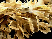

Although zooids are microscopic, colonies range in size from 1 cm (1⁄2 in) to over 1 m (3 ft 3 in).[17] However, the majority are under 10 cm (4 in) across.[20] The shapes of colonies vary widely, depend on the pattern of budding by which they grow, the variety of zooids present and the type and amount of skeletal material they secrete.[17]
Some marine species are bush-like or fan-like, supported by "trunks" and "branches" formed by kenozooids, with feeding autozooids growing from these. Colonies of these types are generally unmineralized but may have exoskeletons made of chitin.[17] Others look like small corals, producing heavy lime skeletons.[43] Many species form colonies which consist of sheets of autozooids. These sheets may form leaves, tufts or, in the genus Thalamoporella, structures that resemble an open head of lettuce.[17]
The most common marine form, however, is encrusting, in which a one-layer sheet of zooids spreads over a hard surface or over seaweed. Some encrusting colonies may grow to over 50 cm (1 ft 8 in) and contain about 2,000,000 zooids.[17] These species generally have exoskeletons reinforced with calcium carbonate, and the openings through which the lophophores protrude are on the top or outer surface.[17] The moss-like appearance of encrusting colonies is responsible for the phylum's name (Ancient Greek words βρύον brúon meaning 'moss' and ζῷον zôion meaning 'animal').[44] Large colonies of encrusting species often have "chimneys", gaps in the canopy of lophophores, through which they swiftly expel water that has been sieved, and thus avoid re-filtering water that is already exhausted.[45] They are formed by patches of non-feeding heterozooids.[46] New chimneys appear near the edges of expanding colonies, at points where the speed of the outflow is already high, and do not change position if the water flow changes.[47]
Some freshwater species secrete a mass of gelatinous material, up to 1 m (3 ft 3 in) in diameter, to which the zooids stick. Other freshwater species have plant-like shapes with "trunks" and "branches", which may stand erect or spread over the surface. A few species can creep at about 2 cm (3⁄4 in) per day.[17]
Each colony grows by asexual budding from a single zooid known as the ancestrula,[17] which is round rather than shaped like a normal zooid.[20] This occurs at the tips of "trunks" or "branches" in forms that have this structure. Encrusting colonies grow round their edges. In species with calcareous exoskeletons, these do not mineralize until the zooids are fully grown. Colony lifespans range from one to about 12 years, and the short-lived species pass through several generations in one season.[17]
Species that produce defensive zooids do so only when threats have already appeared, and may do so within 48 hours.[27] The theory of "induced defenses" suggests that production of defenses is expensive and that colonies which defend themselves too early or too heavily will have reduced growth rates and lifespans. This "last minute" approach to defense is feasible because the loss of zooids to a single attack is unlikely to be significant.[27] Colonies of some encrusting species also produce special heterozooids to limit the expansion of other encrusting organisms, especially other bryozoans. In some cases this response is more belligerent if the opposition is smaller, which suggests that zooids on the edge of a colony can somehow sense the size of the opponent. Some species consistently prevail against certain others, but most turf wars are indecisive and the combatants soon turn to growing in uncontested areas.[27] Bryozoans competing for territory do not use the sophisticated techniques employed by sponges or corals, possibly because the shortness of bryozoan lifespans makes heavy investment in turf wars unprofitable.[27]
Bryozoans have contributed to carbonate sedimentation in marine life since the Ordovician period. Bryozoans take responsibility for many of the colony forms, which have evolved in different taxonomic groups and vary in sediment producing ability. The nine basic bryozoan colony-forms include: encrusting, dome-shaped, palmate, foliose, fenestrate, robust branching, delicate branching, articulated and free-living. Most of these sediments come from two distinct groups of colonies: domal, delicate branching, robust branching and palmate; and fenestrate. Fenestrate colonies generate rough particles both as sediment and components of stromatoporoids coral reefs. The delicate colonies however, create both coarse sediment and form the cores of deep-water, subphotic biogenic mounds. Nearly all post- bryozoan sediments are made up of growth forms, with the addition to free-living colonies which include significant numbers of various colonies. "In contrast to the Palaeozoic, post-Palaeozoic bryozoans generated sediment varying more widely with the size of their grains; they grow as they moved from mud, to sand, to gravel."[48]
Taxonomy
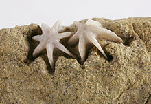
The phylum was originally called "Polyzoa", but this name was eventually replaced by Ehrenberg's term "Bryozoa".[13][49][50] The name "Bryozoa" was originally applied only to the animals also known as Ectoprocta (lit. 'outside-anus'),[51] in which the anus lies outside the "crown" of tentacles. After the discovery of the Entoprocta (lit. 'inside-anus'),[52] in which the anus lies within a "crown" of tentacles, the name "Bryozoa" was promoted to phylum level to include the two classes Ectoprocta and Entoprocta.[53] However, in 1869 Hinrich Nitsche regarded the two groups as quite distinct for a variety of reasons, and coined the name "Ectoprocta" for Ehrenberg's "Bryozoa".[5][54] Despite their apparently similar methods of feeding, they differed markedly anatomically; in addition to the different positions of the anus, ectoprocts have hollow tentacles and a coelom, while entoprocts have solid tentacles and no coelom. Hence the two groups are now widely regarded as separate phyla, and the name "Bryozoa" is now synonymous with "Ectoprocta".[53] This has remained the majority view ever since, although most publications have preferred the name "Bryozoa" rather than "Ectoprocta".[50] Nevertheless, some notable scientists have continued to regard the "Ectoprocta" and Entoprocta as close relatives and group them under "Bryozoa".[54]
The ambiguity about the scope of the name "Bryozoa" led to proposals in the 1960s and 1970s that it should be avoided and the unambiguous term "Ectoprocta" should be used.[55] However, the change would have made it harder to find older works in which the phylum was called "Bryozoa", and the desire to avoid ambiguity, if applied consistently to all classifications, would have necessitated renaming of several other phyla and many lower-level groups.[49] In practice, zoological naming of split or merged groups of animals is complex and not completely consistent.[56] Works since 2000 have used various names to resolve the ambiguity, including: "Bryozoa",[17][20] "Ectoprocta",[23][27] "Bryozoa (Ectoprocta)",[30] and "Ectoprocta (Bryozoa)".[57] Some have used more than one approach in the same work.[58]
The common name "moss animals" is the literal meaning of "Bryozoa", from Greek βρυόν ('moss') and ζῷα ('animals'), based on the mossy appearance of encrusting species.[59]
Until 2008 there were "inadequately known and misunderstood type species belonging to the Cyclostome Bryozoan family Oncousoeciidae."[60] Modern research and experiments have been done using low-vacuum scanning electron microscopy of uncoated type material to critically examine and perhaps revise the taxonomy of three genera belonging to this family, including Oncousoecia, Microeciella, and Eurystrotos. This method permits data to be obtained that would be difficult to recognize with an optical microscope. The valid type species of Oncousoecia was found to be Oncousoecia lobulata. This interpretation stabilizes Oncousoecia by establishing a type species that corresponds to the general usage of the genus. Fellow Oncousoeciid Eurystrotos is now believed to be not conspecific with O. lobulata, as previously suggested, but shows enough similarities to be considered a junior synonym of Oncousoecia. Microeciella suborbicularus has also been recently distinguished from O. lobulata and O. dilatans, using this modern method of low vacuum scanning, with which it has been inaccurately synonymized with in the past. A new genus has also been recently discovered called Junerossia in the family Stomachetosellidae, along with 10 relatively new species of bryozoa such as Alderina flaventa, Corbulella extenuata, Puellina septemcryptica, Junerossia copiosa, Calyptotheca kapaaensis, Bryopesanser serratus, Cribellopora souleorum, Metacleidochasma verrucosa, Disporella compta, and Favosipora adunca.[61]
Classification and diversity
Counts of formally described species range between 4,000 and 4,500.[62] The Gymnolaemata and especially Cheilostomata have the greatest numbers of species, possibly because of their wide range of specialist zooids.[27] Under the Linnaean system of classification, which is still used as a convenient way to label groups of organisms,[63] living members of the phylum Bryozoa are divided into:[17][27]
| Class | Phylactolaemata | Stenolaemata | Gymnolaemata | |
|---|---|---|---|---|
| Order | Plumatellida[64] | Cyclostomatida | Ctenostomatida | Cheilostomata |
| Environments | Freshwater | Marine | Mostly marine | |
| Lip-like epistome overhanging mouth | Yes | none | ||
| Colony shapes | Gelatinous masses or tubular branching structures[65] | Erect or encrusting[66] | Erect, encrusting or free-living | |
| Exoskeleton material | Gelatinous or membranous; unmineralized | Mineralized | Chitin, gelatinous or membranous; unmineralized | Mineralized |
| Operculum ("lid") | none | none[66] (except in family Eleidae)[67] | None in most species | Yes (except in genus Bugula) |
| Shape of lophophore | U-shaped appearance(except in genus Fredericella, whose lophophore is circular) | Circular | ||
| How lophophore extended | Compressing the whole body wall | Compressing the membranous sac(separate inner layer of epithelium that lines the coelom) | Compressing the whole body wall | Pulling inwards of a flexible section of body wall, or making an internal sac expand. |
| Types of zooid | Autozooids only | Limited heterozooids, mainly gonozooids[68] | Stolons and spines as well as autozooids[68] | Full range of types |
Fossil record
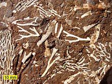
| Stereo image | |||
|---|---|---|---|
| |||
| |||
| |||
| |||
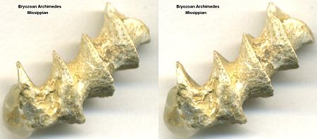
| Fossilized skeleton of Archimedes Bryozoan | |||
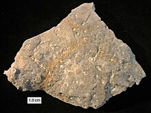
Fossils of about 15,000 bryozoan species have been found. Bryozoans are among the three dominant groups of Paleozoic fossils.[69] Bryozoans with calcitic skeletons were a major source of the carbonate minerals that make up limestones, and their fossils are incredibly common in marine sediments worldwide from the Ordovician onward. However, unlike corals and other colonial animals found in the fossil record, Bryozoan colonies did not reach large sizes.[70] Fossil bryozoan colonies are typically found highly fragmented and scattered; the preservation of complete zoaria is uncommon in the fossil record, and relatively little study has been devoted to reassembling fragmented zoaria.[71] The largest known fossil colonies are branching trepostome bryozoans from Ordovician rocks in the United States, reaching 66 centimeters in height.[70]
The oldest species with a mineralized skeleton occurs in the Lower Ordovician.[1] It is likely that the first bryozoans appeared much earlier and were entirely soft-bodied, and the Ordovician fossils record the appearance of mineralized skeletons in this phylum.[5] By the Arenigian stage of the Early Ordovician period,[20][72] about 480 million years ago, all the modern orders of stenolaemates were present,[73] and the ctenostome order of gymnolaemates had appeared by the Middle Ordovician, about 465 million years ago. The Early Ordovician fossils may also represent forms that had already become significantly different from the original members of the phylum.[73] Ctenostomes with phosphatized soft tissue are known from the Devonian.[74] Other types of filter feeders appeared around the same time, which suggests that some change made the environment more favorable for this lifestyle.[20] Fossils of cheilostomates, an order of gymnolaemates with mineralized skeletons, first appear in the Mid Jurassic, about 172 million years ago, and these have been the most abundant and diverse bryozoans from the Cretaceous to the present.[20] Evidence compiled from the last 100 million years show that cheilostomatids consistently grew over cyclostomatids in territorial struggles, which may help to explain how cheilostomatids replaced cyclostomatids as the dominant marine bryozoans.[75] Marine fossils from the Paleozoic era, which ended 251 million years ago, are mainly of erect forms, those from the Mesozoic are fairly equally divided by erect and encrusting forms, and more recent ones are predominantly encrusting.[76] Fossils of the soft, freshwater phylactolaemates are very rare,[20] appear in and after the Late Permian (which began about 260 million years ago) and consist entirely of their durable statoblasts.[65] There are no known fossils of freshwater members of other classes.[65]
Evolutionary family tree

Scientists are divided about whether the Bryozoa (Ectoprocta) are a monophyletic group (whether they include all and only a single ancestor species and all its descendants), about what are the phylum's closest relatives in the family tree of animals, and even about whether they should be regarded as members of the protostomes or deuterostomes, the two major groups that account for all moderately complex animals.
Molecular phylogeny, which attempts to work out the evolutionary family tree of organisms by comparing their biochemistry and especially their genes, has done much to clarify the relationships between the better-known invertebrate phyla.[53] However, the shortage of genetic data about "minor phyla" such as bryozoans and entoprocts has left their relationships to other groups unclear.[54]
Traditional view
The traditional view is that the Bryozoa are a monophyletic group, in which the class Phylactolaemata is most closely related to Stenolaemata and Ctenostomatida, the classes that appear earliest in the fossil record.[77] However, in 2005 a molecular phylogeny study that focused on phylactolaemates concluded that these are more closely related to the phylum Phoronida, and especially to the only phoronid species that is colonial, than they are to the other ectoproct classes. That implies that the Entoprocta are not monophyletic, as the Phoronida are a sub-group of ectoprocts but the standard definition of Entoprocta excludes the Phoronida.[77]
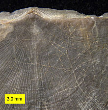
In 2009 another molecular phylogeny study, using a combination of genes from mitochondria and the cell nucleus, concluded that Bryozoa is a monophyletic phylum, in other words includes all the descendants of a common ancestor that is itself a bryozoan. The analysis also concluded that the classes Phylactolaemata, Stenolaemata and Gymnolaemata are also monophyletic, but could not determine whether Stenolaemata are more closely related to Phylactolaemata or Gymnolaemata. The Gymnolaemata are traditionally divided into the soft-bodied Ctenostomatida and mineralized Cheilostomata, but the 2009 analysis considered it more likely that neither of these orders is monophyletic and that mineralized skeletons probably evolved more than once within the early Gymnolaemata.[5]
Bryozoans' relationships with other phyla are uncertain and controversial. Traditional phylogeny, based on anatomy and on the development of the adult forms from embryos, has produced no enduring consensus about the position of ectoprocts.[23] Attempts to reconstruct the family tree of animals have largely ignored ectoprocts and other "minor phyla", which have received little scientific study because they are generally tiny, have relatively simple body plans, and have little impact on human economies – despite the fact that the "minor phyla" include most of the variety in the evolutionary history of animals.[79]
In the opinion of Ruth Dewel, Judith Winston, and Frank McKinney, "Our standard interpretation of bryozoan morphology and embryology is a construct resulting from over 100 years of attempts to synthesize a single framework for all invertebrates," and takes little account of some peculiar features of ectoprocts.[73]

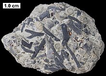
In ectoprocts, all of the larva's internal organs are destroyed during the metamorphosis to the adult form and the adult's organs are built from the larva's epidermis and mesoderm, while in other bilaterians some organs including the gut are built from endoderm. In most bilaterian embryos the blastopore, a dent in the outer wall, deepens to become the larva's gut, but in ectoprocts the blastopore disappears and a new dent becomes the point from which the gut grows. The ectoproct coelom is formed by neither of the processes used by other bilaterians, enterocoely, in which pouches that form on the wall of the gut become separate cavities, nor schizocoely, in which the tissue between the gut and the body wall splits, forming paired cavities.[73]
Entoprocts
When entoprocts were discovered in the 19th century, they and bryozoans (ectoprocts) were regarded as classes within the phylum Bryozoa, because both groups were sessile animals that filter-fed by means of a crown of tentacles that bore cilia.
From 1869 onwards increasing awareness of differences, including the position of the entoproct anus inside the feeding structure and the difference in the early pattern of division of cells in their embryos, caused scientists to regard the two groups as separate phyla,[54] and "Bryozoa" became just an alternative name for ectoprocts, in which the anus is outside the feeding organ.[53] A series of molecular phylogeny studies from 1996 to 2006 have also concluded that bryozoans (ectoprocts) and entoprocts are not sister groups.[54]
However, two well-known zoologists, Claus Nielsen and Thomas Cavalier-Smith, maintain on anatomical and developmental grounds that bryozoans and entoprocts are member of the same phylum, Bryozoa. A molecular phylogeny study in 2007 also supported this old idea, while its conclusions about other phyla agreed with those of several other analyses.[54]
Grouping into the Lophophorata
By 1891 bryozoans (ectoprocts) were grouped with phoronids in a super-phylum called "Tentaculata". In the 1970s comparisons between phoronid larvae and the cyphonautes larva of some gymnolaete bryozoans produced suggestions that the bryozoans, most of which are colonial, evolved from a semi-colonial species of phoronid.[80] Brachiopods were also assigned to the "Tentaculata", which were renamed Lophophorata as they all use a lophophore for filter feeding.[53]
The majority of scientists accept this,[53] but Claus Nielsen thinks these similarities are superficial.[23] The Lophophorata are usually defined as animals with a lophophore, a three-part coelom and a U-shaped gut.[80] In Nielsen's opinion, phoronids' and brachiopods' lophophores are more like those of pterobranchs,[23] which are members of the phylum Hemichordata.[81] Bryozoan's tentacles bear cells with multiple cilia, while the corresponding cells of phoronids', brachiopods' and pterobranchs' lophophores have one cilium per cell; and bryozoan tentacles have no hemal canal ("blood vessel"), which those of the other three phyla have.[23]
If the grouping of bryozoans with phoronids and brachiopods into Lophophorata is correct, the next issue is whether the Lophophorata are protostomes, along with most invertebrate phyla, or deuterostomes, along with chordates, hemichordates and echinoderms.
The traditional view was that lophophorates were a mix of protostome and deuterostome features. Research from the 1970s onwards suggested they were deuterostomes, because of some features that were thought characteristic of deuterostomes: a three-part coelom; radial rather than spiral cleavage in the development of the embryo;[53] and formation of the coelom by enterocoely.[23] However the coelom of ectoproct larvae shows no sign of division into three sections,[80] and that of adult ectoprocts is different from that of other coelomate phyla as it is built anew from epidermis and mesoderm after metamorphosis has destroyed the larval coelom.[73]
Lophophorate molecular phylogenetics
Molecular phylogeny analyses from 1995 onwards, using a variety of biochemical evidence and analytical techniques, placed the lophophorates as protostomes and closely related to annelids and molluscs in a super-phylum called Lophotrochozoa.[53][82] "Total evidence" analyses, which used both morphological features and a relatively small set of genes, came to various conclusions, mostly favoring a close relationship between lophophorates and Lophotrochozoa.[82] A study in 2008, using a larger set of genes, concluded that the lophophorates were closer to the Lophotrochozoa than to deuterostomes, but also that the lophophorates were not monophyletic. Instead, it concluded that brachiopods and phoronids formed a monophyletic group, but bryozoans (ectoprocts) were closest to entoprocts, supporting the original definition of "Bryozoa".[82]
They are the only major phylum of exclusively clonal animals, composed of modular units known as zooids. Because they thrive in colonies, colonial growth allows them to develop unrestricted variations in form. Despite this, only a small number of basic growth forms have been found and have commonly reappeared throughout the history of the bryozoa.[69]

